File:3D-fluorescence imaging for high throughput analysis of microbial eukaryotes (b left).jpg

Original file(1,982 × 1,918 pixels, file size: 167 KB, MIME type: image/jpeg)

This file is from Wikimedia Commons and may be used by other projects. The description on its file description page there is shown below.

Summary

Description
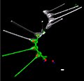
English: Environmental high-content fluorescence microscopy (e-HCFM): automated, 3D, and multichannel imaging for planktonic micro-eukaryotes: a chain of diatoms (Asterionellopsis sp., Heterokonta). Scale bar 5 µm.
Date
Source
image extraction process
This file has been extracted from another file
: 3D-fluorescence imaging for high throughput analysis of microbial eukaryotes.jpg
original file
Original: doi:10.7554/eLife.26066 Quantitative 3D-imaging for cell biology and ecology of environmental microbial eukaryotes, Fig. 1b (left panel)
Author Sebastien Colin, Luis Pedro Coelho, Shinichi Sunagawa, Chris Bowler, Eric Karsenti, Peer Bork, Rainer Pepperkok, Colomban de Vargas
Other versions
image extraction process
This file has been extracted from another file
: 3D-fluorescence imaging for high throughput analysis of microbial eukaryotes.jpg
original file

Licensing

w:en:Creative Commons
attribution share alike
This file is licensed under the Creative Commons Attribution-Share Alike 4.0 International license.
You are free:
  • to share – to copy, distribute and transmit the work
  • to remix – to adapt the work
Under the following conditions:
  • attribution – You must give appropriate credit, provide a link to the license, and indicate if changes were made. You may do so in any reasonable manner, but not in any way that suggests the licensor endorses you or your use.
  • share alike – If you remix, transform, or build upon the material, you must distribute your contributions under the same or compatible license as the original.

Captions

Add a one-line explanation of what this file represents

Items portrayed in this file

depicts

26 September 2017

image/jpeg

4a0f67dd039681b82024285ad15933d047f7fd11

171,325 byte

1,918 pixel

1,982 pixel

File history

Click on a date/time to view the file as it appeared at that time.

Date/TimeThumbnailDimensionsUserComment
current10:29, 1 April 2022Thumbnail for version as of 10:29, 1 April 20221,982 × 1,918 (167 KB)ErnstsUploaded a work by Sebastien Colin, Luis Pedro Coelho, Shinichi Sunagawa, Chris Bowler, Eric Karsenti, Peer Bork, Rainer Pepperkok, Colomban de Vargas from Cropped from File:3D-fluorescence imaging for high throughput analysis of microbial eukaryotes.jpg. Original: doi:10.7554/eLife.26066 Quantitative 3D-imaging for cell biology and ecology of environmental microbial eukaryotes, Fig. 1b (left panel) with UploadWizard

The following page uses this file:

Global file usage

The following other wikis use this file: